4.9 Critical joints discussed in this course
4.9.1 The hip

@Bartel2006
- The hip is a synovial ball and socket joint.
- The head of the femur rotates relative to the acetabulum of the pelvis
- Contact can (sometimes) be modeled as a single force acting
through the center of the joint
- This approximation isn’t perfect
@Bartel2006
- Modeling the hip
- We might model the femoral diaphysis as a hollow circular beam
- The metaphysis can be modeled as an elastic or rigid link, or modeled using FEA if detailed outcome is desired
- Contact in the ball and socket leads to complex load distributions
@Bartel2006
- Muscle attachments to the greater and lesser trochanters
and bony ridges such as the linea aspera
- These allow for a greater moment arm for the muscles
4.9.2 The knee

@Blausen2014
- A bicondylar synovial joint which allows the femur and tibia to rotate, twist, and slide relative to each other
- Each motion is necessary… otherwise abnormal forces develop and cause rapid deterioration of the joint

@Bartel2006
- Important structures include:
- Medial collateral ligament (MCL)
- Lateral collateral ligament (LCL)
- Quadriceps tendon
- Patellar ligament
- Anterior Cruciate ligament (ACL)
- Posterior Cruciate Ligament (PCL)
- Menisci (two crescent-shaped pads that help distribute the loads from the femoral condyles to the tibial plateaus)
- ACL and PCL are within the knee joint capsule and pass through the notch between the condyles, the others are outside the capsule
4.9.3 Spine

@Bartel2006


OpenStax College CCSA3.0
“Cartilaginous” type joint
Connective tissue is a fibrocartilage structure called the Intervertebral disc
Healthy disk acts like a tire… needs to have sufficient fluid inside.
- Aging causes the gel-like fluid to solidify with negative consequences
- Largest avascular tissue in the body
4.9.4 Anterior portion of the intervertebral joint
- Pedicles and laminae form an arch – the vertebral foramen (hole) for the spinal cord


Dr.foksha CCSA4.0
- Adjacent vertebrae are connected through articulating joints (called facets) that constrain the motion between the vertebrae and limit twisting and extension of the spinal column
- The structure supports the upper body and protects the spinal cord.
4.9.5 Vertebral attachments
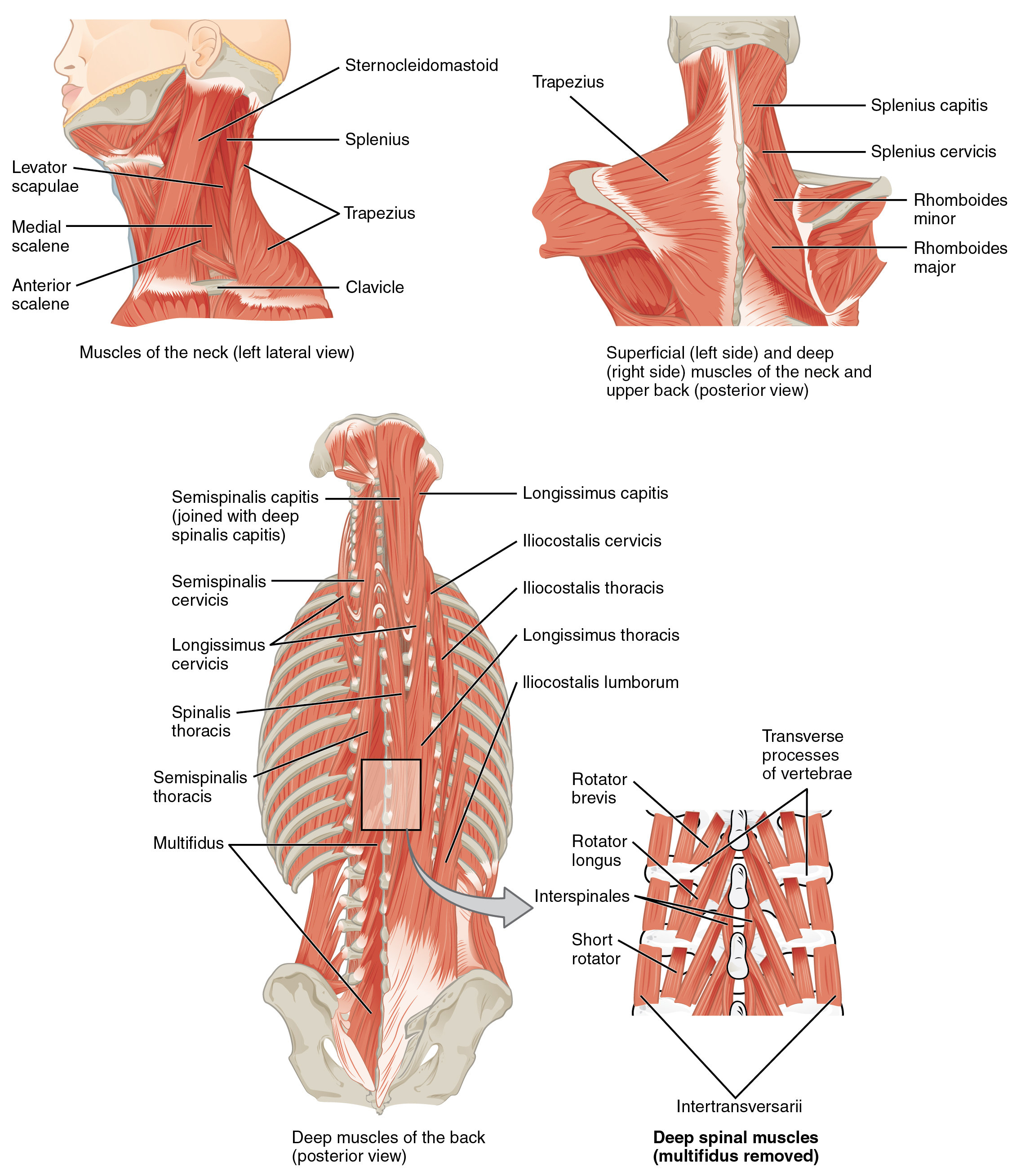
OpenStax College CCSA3.0
- Back muscles attach \(\approx 5\)cm posterior to the center of the vertebral body
- Center of sagittal bending is approximately within the disk
Latissimus dorsi

Anatomography CCSA2.1 Japan

@Bartel2006