4.6 Bones
4.6.1 Major boney structures examined in this course

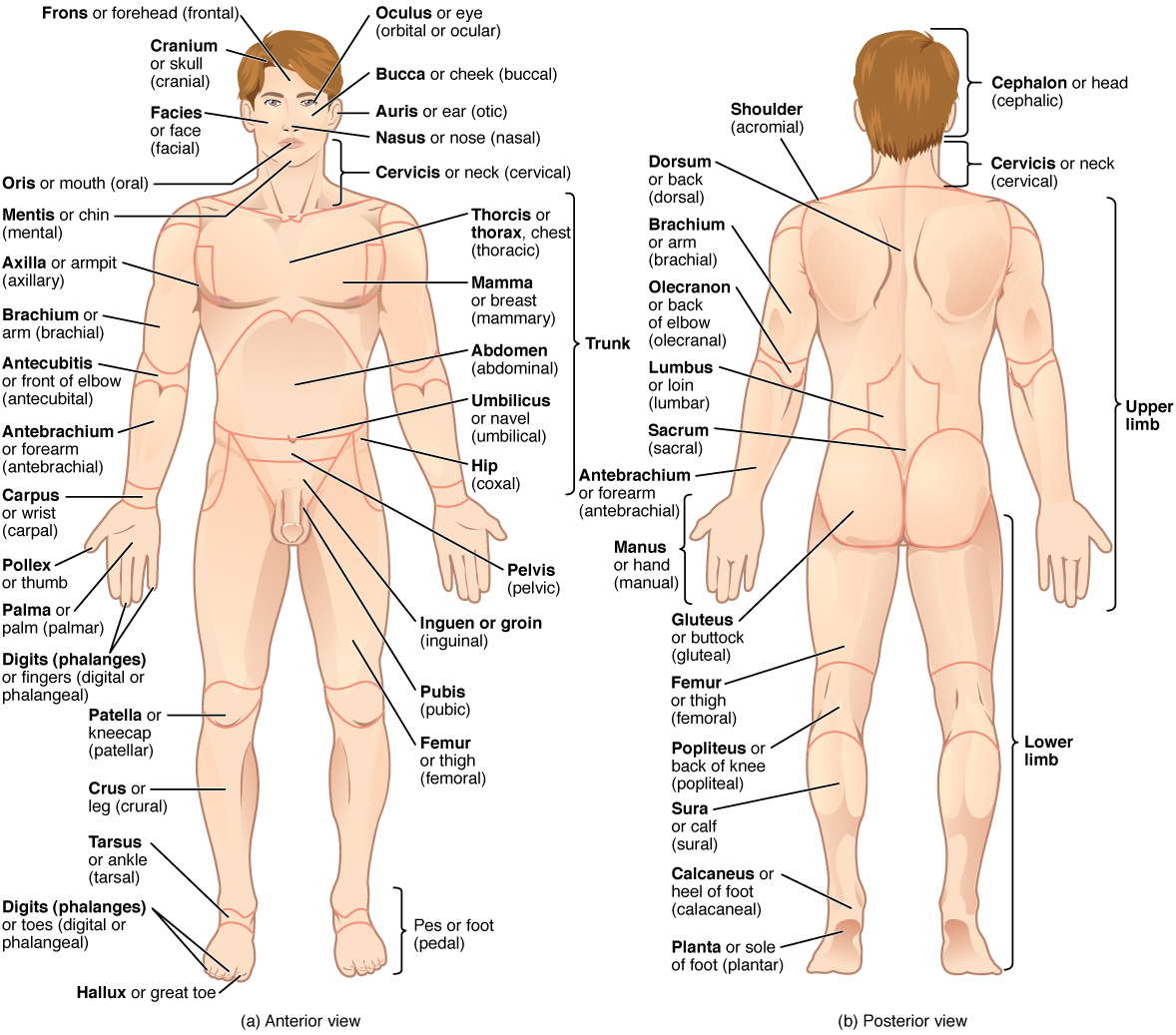
The human body is shown in anatomical position in an (a) anterior view and a (b) posterior view. The regions of the body are labeled in boldface.
- There are typically 206 bones in the human body (+4 sesamoid bones in the foot)
- Can be categorized into roughly 4 groups
- Long bones (femur, tibia, humerus, etc)
- Long in one direction, tubular cross sections
- Short bones (wrist, ankle, hand, foot, etc)
- Have similar dimensions in all directions
- Flat bones (scapula, skull, pelvis, etc)
- Are short in one dimension relative to the others
- Irregular
- Those that don’t easily fit into the other categories
- Long bones (femur, tibia, humerus, etc)
4.6.2 Bone tissue types
Femoral head with metastasis
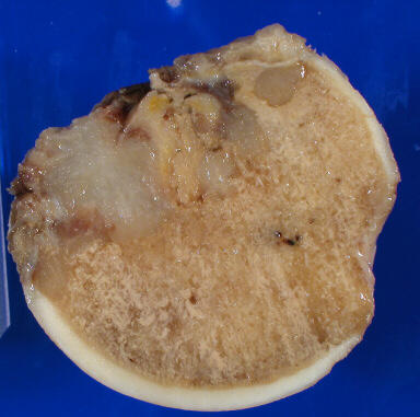
Femoral_head_with_bone_metastasis
- There are two types of bone tissue
- Cortical (compact) bone
- Dense, stiff, strong
- Load carrying bone
- Cancellous (trabecular) bone
- Less dense, less stiff
- What functions does it serve?
- Cortical (compact) bone
4.6.3 Long bone

@Bartel2006
The shaft (diaphysis) grows from its ends at growth plates (physis)
- The shaft has a medullary cavity filled with yellow marrow (fat and primitive blood cells)
- The proximal humerus and femur have red marrow within their cancellous bone tissue. (Red blood cells are made here)
- The medullary cavity serves no structural purpose in normal bone
- Implants often interface with the medullary cavity as a means of attachment (ie intramedullary nail)
- The epiphysis (bone end) grows from separate ossification centers at the end of the bone
- Metaphysis is the region between the diaphysis and the epiphysis
4.6.4 Anatomy of a Long Bone

@OpenStaxAnatomy2020 Ch. 6
A typical long bone shows the gross anatomical characteristics of bone.
(slide credit: @OpenStaxAnatomy2020 Ch. 6)
4.6.5 Periosteum and Endosteum

@OpenStaxAnatomy2020 Ch. 6
The periosteum forms the outer surface of bone, and the endosteum lines the medullary cavity.
(slide credit: @OpenStaxAnatomy2020 Ch. 6)
4.6.6 Anatomy of a Flat Bone

@OpenStaxAnatomy2020 Ch. 6
This cross-section of a flat bone shows the spongy bone (diploë) lined on either side by a layer of compact bone.
(slide credit: @OpenStaxAnatomy2020 Ch. 6)
4.6.7 Bone Features

@OpenStaxAnatomy2020 Ch. 6
The surface features of bones depend on their function, location, attachment of ligaments and tendons, or the penetration of blood vessels and nerves.
(slide credit: @OpenStaxAnatomy2020 Ch. 6)