4.7 Joints
4.7.0.1 Joints can be classified functionally or structurally
- Functional classification depends on the amount of
relative motion… less convenient
- Large motions – diarthrodial joint
- Structural classification depends on joint mechanism
4.7.1 Structural classification of joints
- Fibrous (or Immovable) joints
- Held together by a thin layer of strong connective tissue
- No movement between the bones such
- Sutures of the skull
- Teeth in their sockets
- Cartilaginous joints
- Bones attached by white fibrocartilaginous discs and ligaments
- Limited degree of movement
- Intervertebral discs
- The cartilage in the symphysis (binds the pubic bones together at the front of the pelvic girdle)
- The cartilage between the sacrum and the hip bone
- Synovial joints (subset of diarthrodial joints)
- Covered with a layer of smooth articular (hyaline) cartilage
- Enclosed by a bag-like capsular ligament which constrains the joint and helps contain the synovial fluid
- The capsular ligament is lined with a synovial membrane. This membrane secretes synovial fluid into the cavity as a lubricant
- In addition to the capsule, the bones are constrained by other strong ligaments
- Very low friction coefficient
- human knee – \(\mu=0.005-0.02\)
- human hip – \(\mu=0.01-0.04\)
- Lower than most engineered structures
4.7.2 Synovial joints
@Bartel2006
- Ball and socket
- Condylar joints
- Multiple bone joints (Compound Joints)
4.7.3 Ball and socket
- The rounded head interfaces the cup-shaped socket of another
- Hip
- Femoral head (ball) and acetabulum (socket)
- Shoulder
- Humeral head (ball) and glenoid (socket)
- The glenoid is nearly flat… thus the shoulder requires significant soft tissue for stabilization
- Hip
4.7.4 Bi-condylar joints
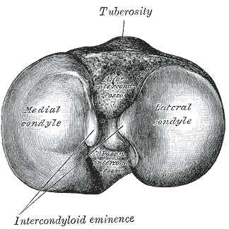
- The condyles are the enlarged regions at knuckles of any
joint.
- They are enlarged, in part, to minimize the stresses of load transfer.
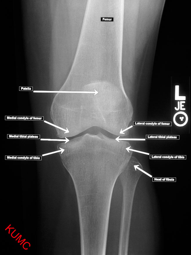
- Examples: medial and lateral condyles of the femur and tibia
4.7.5 Bi-condylar joints
- “Bi” - Pairs of articulating surfaces
- Two curved condyles articulate against relatively flat surfaces of the mating bone
- There is little kinematic constraint, thus, extensive soft tissue is required for stability.

Unlabelled Image Missing
- Having two condyles allows for resistance to moments in planes
perpendicular to the plane of motion.
- The two condyles and their supporting soft tissues only allow
small angular rotations in planes perpendicular to the intended
plane
- Less muscular restraint required about these planes
- The two condyles and their supporting soft tissues only allow
small angular rotations in planes perpendicular to the intended
plane
- These joints have extensive range of motion in one plane
- ie the knee has extensive flexion/extension in the sagittal plane
4.7.7 Multiple bone joints

- More difficult to describe than ball-and-socket or bi-condylar joints
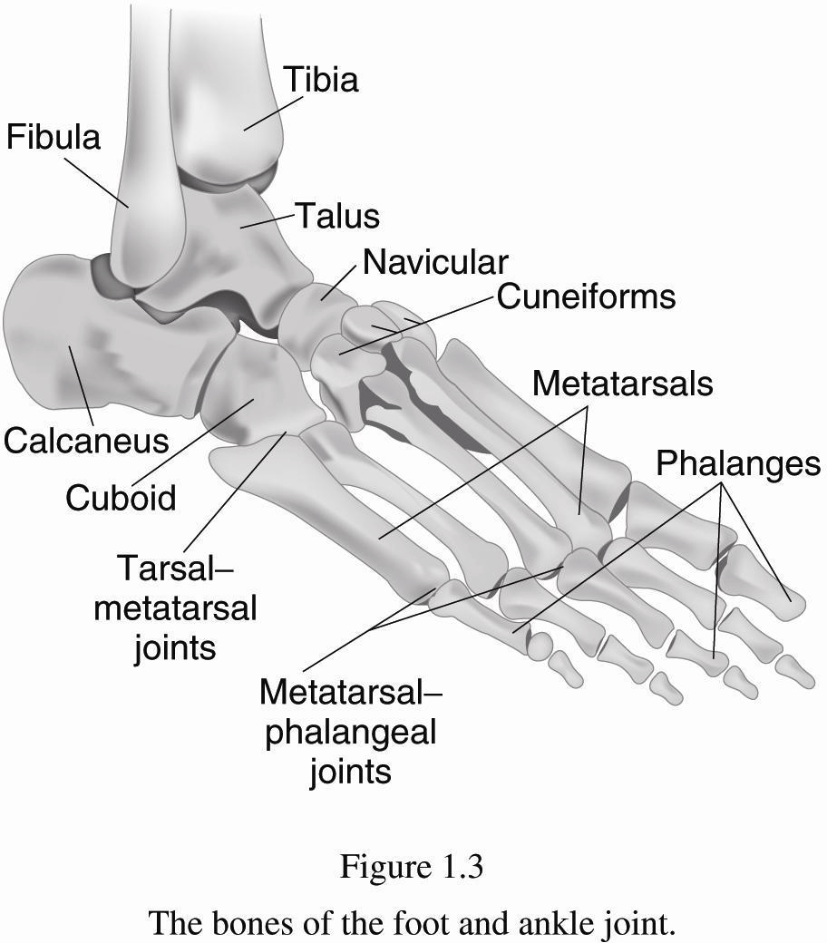
- Examples: wrist (8 carpal bones), ankle (7 tarsal bones)
- Bound by lots of ligaments and have synovial fluid
- Small motions between individual joints add up to larger motions
![AP Hip, AP Shoulder, and Axial Shoulder X-rays ^[http://medical-imaging2012.blogspot.com/2012/02/how-to-take-x-rays.html]](img/uncl/xray_HIP_AP.jpg)
![AP Hip, AP Shoulder, and Axial Shoulder X-rays ^[http://medical-imaging2012.blogspot.com/2012/02/how-to-take-x-rays.html]](img/uncl/xray_shoulder_normal1.jpg)
![AP Hip, AP Shoulder, and Axial Shoulder X-rays ^[http://medical-imaging2012.blogspot.com/2012/02/how-to-take-x-rays.html]](img/uncl/glenoid_axial.jpg)


![AP Hip, Oblique, and Lateral X-rays of the Foot ^[http://orthopedicgallery.com/portfolio/foot-x-ray/]](img/uncl/xray-Foot-AP-150x300.png)
![AP Hip, Oblique, and Lateral X-rays of the Foot ^[http://orthopedicgallery.com/portfolio/foot-x-ray/]](img/uncl/xray-Foot-oblique-165x300.png)
![AP Hip, Oblique, and Lateral X-rays of the Foot ^[http://orthopedicgallery.com/portfolio/foot-x-ray/]](img/uncl/xray-Foot-Lat-300x150.png)