ME5200 - Orthopaedic Biomechanics:
Lecture 2
Anatomy, functions of the musculoskeletal system, joints, soft tissue
Musculoskeletal Anatomy
Body level concepts
Gross and Microscopic Anatomy
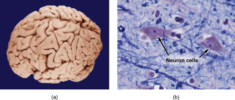
Gross anatomy considers large structures such as the brain.
Microscopic anatomy can deal with the same structures, though at a different scale. This is a micrograph of nerve cells from the brain. LM × 1600.
(slide credit: @OpenStaxAnatomy2020)
Levels of Structural Organization of the Human Body
The organization of the body often is discussed in terms of six distinct levels of increasing complexity, from the smallest chemical building blocks to a unique human organism.
In engineering terms, it is a multi-scale system
(Modified from: @OpenStaxAnatomy2020)
Organ Systems of the Human Body

Organs that work together are grouped into organ systems.
(Modified from: @OpenStaxAnatomy2020)
Anatomical Terms
Directional Terms Applied to the Human Body
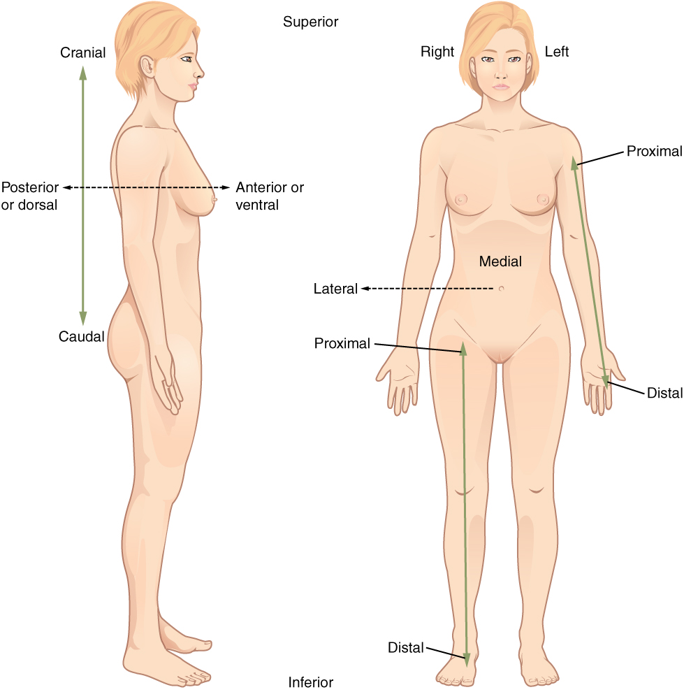
Paired directional terms are shown as applied to the human body.
(slide credit: @OpenStaxAnatomy2020)
Planes of the Body
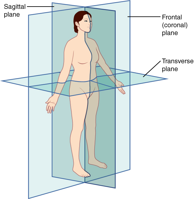
The three planes most commonly used in anatomical and medical imaging are the sagittal, frontal (or coronal), and transverse plane.
(slide credit: @OpenStaxAnatomy2020)
Relative reference terms
- Proximal/Distal aspect
- Nearer to/Further from the origin or attachment point
- Inferior/Superior
- Beneath/Above: Often used in the axial skeleton
- Cranial/Caudal
- Towards/Away from the head
- Lateral/Medial
- Closest to the outside/inside relative to the mid-line in the coronal plane
- Anterior/Posterior
- In front/back (relative position)
- Dorsal/Ventral
- On the back/front
Terms of action
- Flexion/Extension
- The anterior angle between two bones is decreased/increased (except for knees/toes, then posterior angle)
- Abduction/Adduction
- Movement away from/towards the body mid-line in the frontal plane
- Lateral flexion
- Left/right movement of the spine in the frontal plane
- Supination/Pronation
- Rotate the forearm palm forward/backward in the anatomical position
- Dorsiflexion/Plantar flexion
- Rotation of the ankle toes up/down in the sagittal plane
- Inversion/Eversion
- Rotation of the foot up and in/up and out in the frontal plane
- Hyperextension
- Movement beyond anatomical norms
Principal functions of the musculoskeletal system
Critical functions of the bones
- Hematopoiesis (formation of red blood cells)
- Mineral storage (99% of body’s calcium)
- Insufficient calcium intake thus leads to:
- a reduction of calcium storage in the bones and a corresponding reduction the bones’ “mechanical properties”
- an increased fracture risk
- Insufficient calcium intake thus leads to:
- Protection of the vital organs
- Support and motion
Hematopoiesis
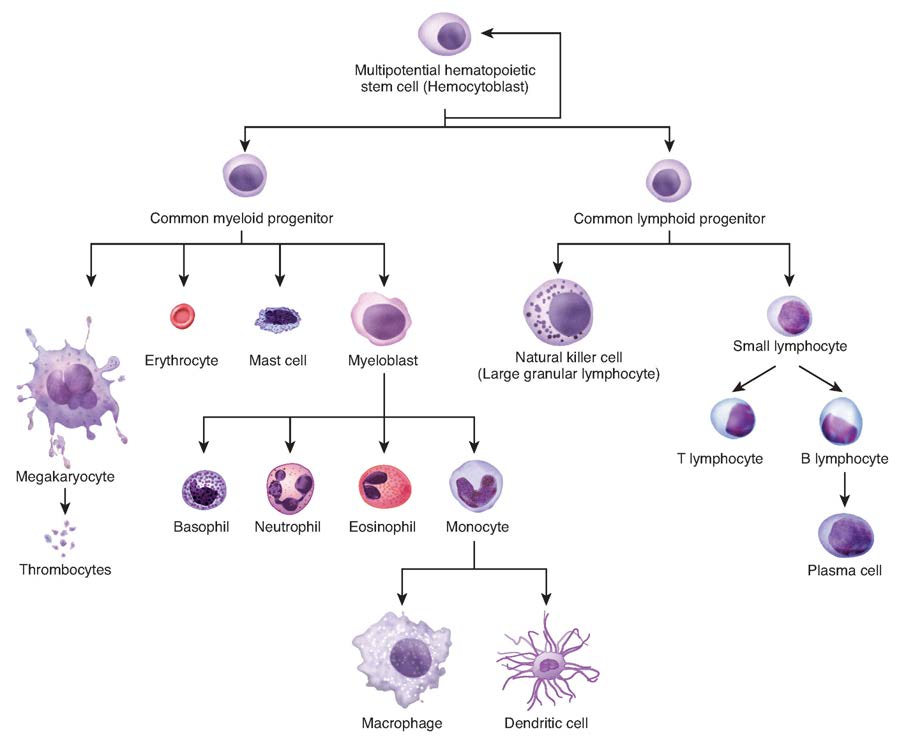
The process of hematopoiesis involves the differentiation of multipotent cells into blood and immune cells. The multipotent hematopoietic stem cells give rise to many different cell types, including the cells of the immune system and red blood cells.
(slide credit: @OpenStaxAnatomy2020 Ch. 3)
Elements of the Human Body
The main elements that compose the human body are shown from most abundant to least abundant.
(slide credit: @OpenStaxAnatomy2020)
Protection of the vital organs
- Distribute loads (stiff cortical bone)
- Absorb energy (softer cancellous bone)
Examples follow:
Bones protect the brain
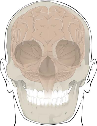
- The cranium completely surrounds and protects the brain from
traumatic injury
- Distributes load and absorbs energy
- Others protective bones
- Ribs, spinal cord, etc
Support and motion
- Provide a framework for force and motion necessary for living
- Bone and joints act as levers, muscles act as actuators
- Muscles apply forces at some moment arm, creating torques to rotate a joint
- Consequences of muscle attachment points (relative to joint position)?
- Disadvantage – loads on joints and muscles are larger than the functional loads applied to the body
- Advantage – small muscle displacements cause large rotations and fast motion
Principal anatomical structures examined to this course
- Bones of the skeleton
- Joints
- Soft tissue
Bones
Major boney structures examined in this course
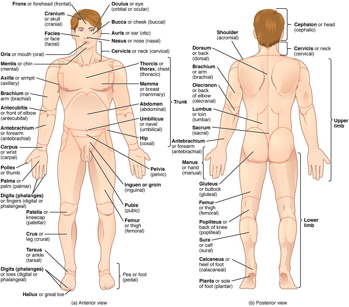
The human body is shown in anatomical position in an (a) anterior view and a (b) posterior view. The regions of the body are labeled in boldface.
- There are typically 206 bones in the human body (+4 sesamoid bones in the foot)
- Can be categorized into roughly 4 groups
- Long bones (femur, tibia, humerus, etc)
- Long in one direction, tubular cross sections
- Short bones (wrist, ankle, hand, foot, etc)
- Have similar dimensions in all directions
- Flat bones (scapula, skull, pelvis, etc)
- Are short in one dimension relative to the others
- Irregular
- Those that don’t easily fit into the other categories
- Long bones (femur, tibia, humerus, etc)
Bone tissue types
Femoral head with metastasis
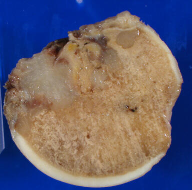
- There are two types of bone tissue
- Cortical (compact) bone
- Dense, stiff, strong
- Load carrying bone
- Cancellous (trabecular) bone
- Less dense, less stiff
- What functions does it serve?
- Cortical (compact) bone
Long bone
The shaft (diaphysis) grows from its ends at growth plates (physis)
- The shaft has a medullary cavity filled with yellow marrow (fat and primitive blood cells)
- The proximal humerus and femur have red marrow within their cancellous bone tissue. (Red blood cells are made here)
- The medullary cavity serves no structural purpose in normal bone
- Implants often interface with the medullary cavity as a means of attachment (ie intramedullary nail)
- The epiphysis (bone end) grows from separate ossification centers at the end of the bone
- Metaphysis is the region between the diaphysis and the epiphysis
Anatomy of a Long Bone

A typical long bone shows the gross anatomical characteristics of bone.
(slide credit: @OpenStaxAnatomy2020 Ch. 6)
Periosteum and Endosteum

The periosteum forms the outer surface of bone, and the endosteum lines the medullary cavity.
(slide credit: @OpenStaxAnatomy2020 Ch. 6)
Anatomy of a Flat Bone

This cross-section of a flat bone shows the spongy bone (diploë) lined on either side by a layer of compact bone.
(slide credit: @OpenStaxAnatomy2020 Ch. 6)
Bone Features

The surface features of bones depend on their function, location, attachment of ligaments and tendons, or the penetration of blood vessels and nerves.
(slide credit: @OpenStaxAnatomy2020 Ch. 6)
Joints
Joints can be classified functionally or structurally
- Functional classification depends on the amount of
relative motion… less convenient
- Large motions – diarthrodial joint
- Structural classification depends on joint mechanism
Structural classification of joints
- Fibrous (or Immovable) joints
- Held together by a thin layer of strong connective tissue
- No movement between the bones such
- Sutures of the skull
- Teeth in their sockets
- Cartilaginous joints
- Bones attached by white fibrocartilaginous discs and ligaments
- Limited degree of movement
- Intervertebral discs
- The cartilage in the symphysis (binds the pubic bones together at the front of the pelvic girdle)
- The cartilage between the sacrum and the hip bone
- Synovial joints (subset of diarthrodial joints)
- Covered with a layer of smooth articular (hyaline) cartilage
- Enclosed by a bag-like capsular ligament which constrains the joint and helps contain the synovial fluid
- The capsular ligament is lined with a synovial membrane. This membrane secretes synovial fluid into the cavity as a lubricant
- In addition to the capsule, the bones are constrained by other strong ligaments
- Very low friction coefficient
- human knee – \(\mu=0.005-0.02\)
- human hip – \(\mu=0.01-0.04\)
- Lower than most engineered structures
Synovial joints
- Ball and socket
- Condylar joints
- Multiple bone joints (Compound Joints)
Ball and socket
- The rounded head interfaces the cup-shaped socket of another
- Hip
- Femoral head (ball) and acetabulum (socket)
- Shoulder
- Humeral head (ball) and glenoid (socket)
- The glenoid is nearly flat… thus the shoulder requires significant soft tissue for stabilization
- Hip
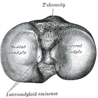
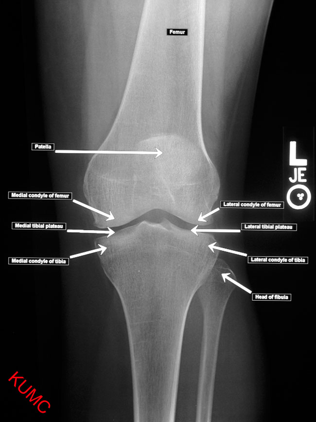
Bi-condylar joints
- The condyles are the enlarged regions at knuckles of any
joint.
- They are enlarged, in part, to minimize the stresses of load transfer.
- Examples: medial and lateral condyles of the femur and tibia
Bi-condylar joints
- “Bi” - Pairs of articulating surfaces
- Two curved condyles articulate against relatively flat surfaces of the mating bone
- There is little kinematic constraint, thus, extensive soft tissue is required for stability.

- Having two condyles allows for resistance to moments in planes
perpendicular to the plane of motion.
- The two condyles and their supporting soft tissues only allow
small angular rotations in planes perpendicular to the intended
plane
- Less muscular restraint required about these planes
- The two condyles and their supporting soft tissues only allow
small angular rotations in planes perpendicular to the intended
plane
- These joints have extensive range of motion in one plane
- ie the knee has extensive flexion/extension in the sagittal plane
Multiple bone joints
Multiple bone joints
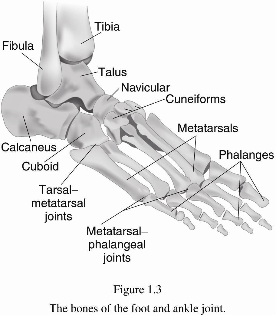
- More difficult to describe than ball-and-socket or bi-condylar joints
- Examples: wrist (8 carpal bones), ankle (7 tarsal bones)
- Bound by lots of ligaments and have synovial fluid
- Small motions between individual joints add up to larger motions
credit a: “WriterHound”/Wikimedia Commons; credit b: Micrograph provided by the Regents of University of Michigan Medical School © 2012↩︎
http://medical-imaging2012.blogspot.com/2012/02/how-to-take-x-rays.html↩︎
https://classes.kumc.edu/som/radanatomy/image.asp?Image=7105-001.jpg&Film=7105&Features=1↩︎
![AP Hip, AP Shoulder, and Axial Shoulder X-rays ^[http://medical-imaging2012.blogspot.com/2012/02/how-to-take-x-rays.html]](img/uncl/xray_HIP_AP.jpg)
![AP Hip, AP Shoulder, and Axial Shoulder X-rays ^[http://medical-imaging2012.blogspot.com/2012/02/how-to-take-x-rays.html]](img/uncl/xray_shoulder_normal1.jpg)
![AP Hip, AP Shoulder, and Axial Shoulder X-rays ^[http://medical-imaging2012.blogspot.com/2012/02/how-to-take-x-rays.html]](img/uncl/glenoid_axial.jpg)
![AP Hip, Oblique, and Lateral X-rays of the Foot ^[http://orthopedicgallery.com/portfolio/foot-x-ray/]](img/uncl/xray-Foot-AP-150x300.png)
![AP Hip, Oblique, and Lateral X-rays of the Foot ^[http://orthopedicgallery.com/portfolio/foot-x-ray/]](img/uncl/xray-Foot-oblique-165x300.png)
![AP Hip, Oblique, and Lateral X-rays of the Foot ^[http://orthopedicgallery.com/portfolio/foot-x-ray/]](img/uncl/xray-Foot-Lat-300x150.png)